
Impacted Wisdom Teeth: Symptoms You Shouldn’t Ignore
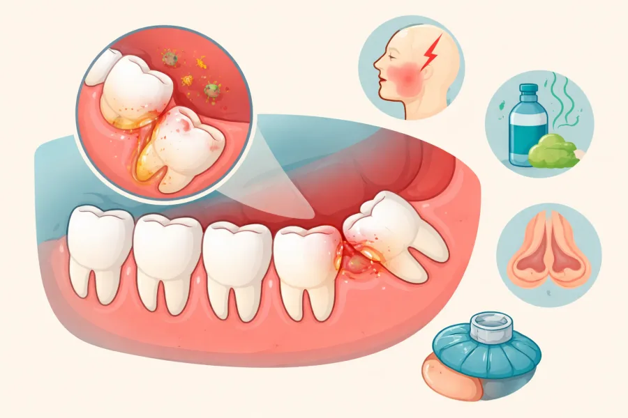
Third molar symptoms rarely begin with unbearable agony. Instead, impacted wisdom teeth usually announce themselves through subtle, easily dismissed warning signs: a persistent stiffness when chewing tough foods, a localized puffiness in the rear gum tissue, or an unexplained sour taste along the back of the tongue. Because these symptoms often ebb and flow over several months, many patients assume the issue has resolved on its own.
Unfortunately, an impacted tooth does not self-correct. When jaw space is insufficient, the tooth remains trapped beneath bone or dense soft tissue, quietly creating chronic inflammation, bacterial reservoirs, and destructive pressure against neighboring teeth. At Oral & Facial Surgery of Miami, Dr. Johanny Caceres helps patients recognize these early clinical signals before minor structural issues escalate into serious dental emergencies.
1. The Anatomy of Impaction: Why Third Molars Become Trapped
Wisdom teeth are the final set of permanent teeth to develop, typically attempting to erupt between the ages of 17 and 25. Anthropological and craniofacial research shows that modern human jaws have evolved to be narrower and shorter than those of our ancestors, leaving the retromolar space severely restricted. By the time the third molars begin their ascent, the dental arch is already fully occupied.
When a tooth lacks the physical corridor required to erupt vertically into a functional bite, it becomes impacted. Depending on the path of embryonic development and surrounding bone architecture, wisdom teeth can become trapped in several distinct orientations, each presenting unique clinical risks.
| Impaction Pattern | Anatomical Orientation | Primary Clinical Risk |
|---|---|---|
| Mesial Impaction | The crown tilts forward at an angle directly into the backside of the adjacent second molar. | Severe food impaction, enamel demineralization, and irreversible root resorption of the second molar. |
| Horizontal Impaction | The tooth lies completely sideways on a 90-degree angle, parallel to the jawline. | Exerts intense transverse pressure against the roots of neighboring teeth and risks cystic bone expansion. |
| Vertical Impaction | The tooth sits upright in a normal orientation but remains blocked beneath the jawbone or thick fibrous gum tissue. | Pericoronitis, recurrent gingival abscesses, and deep periodontal pocket formation along the rear jaw. |
| Distal Impaction | The tooth tilts backward toward the ramus of the mandible or the maxillary tuberosity. | High risk of localized bone loss and sinus floor perforation in the upper jaw. |
2. Warning Signs You Must Never Ignore
While an impacted tooth buried deeply under dense cortical bone may remain silent for a period of time, partially erupted or actively migrating teeth produce recognizable clinical symptoms that signal urgent evaluation is required:
Recurrent Pericoronitis (Swollen Operculum): When a tooth partially pierces the gum, a small flap of mucosal tissue called an operculum remains draped over the biting surface. This creates an ideal, warm incubator for anaerobic oral bacteria and microscopic food particles that cannot be cleansed with a standard toothbrush. The resulting localized flare-up causes severe tenderness, bleeding, and localized throbbing.
Persistent Halitosis and Metallic Aftertaste: Chronic bacterial colonization beneath impacted gum flaps produces foul volatile sulfur compounds. If you notice persistent bad breath or a bitter, metallic taste that brushing, flossing, and therapeutic mouthwashes cannot eradicate, subgingival bacterial activity around a third molar is often the root cause.
Jaw Stiffness and Trismus: When inflammation spreads from the tooth socket into the adjacent masseter and internal pterygoid muscles, opening your mouth comfortably becomes painful or mechanically restricted. Severe trismus (inability to insert two or three fingers vertically between your front teeth) is a classic indicator that the infection is tracking into deeper fascial spaces.
Referred Facial Pain, Headaches, and Sinus Fullness: Because the trigeminal nerve supplies sensory branches throughout your face, jaw, ears, and temples, localized pressure from an impacted lower molar often masquerades as deep earaches or dull temporal tension headaches. Similarly, impacted upper wisdom teeth pushing against the floor of the maxillary sinus can produce chronic nasal congestion and midface pressure often mistaken for seasonal allergies.
3. The Hidden Hazards of Postponing Treatment
Waiting until an impacted wisdom tooth causes excruciating pain often means structural damage has already occurred beneath the gumline. Proactive surgical removal is designed to prevent irreversible damage to tissues and teeth that are meant to last a lifetime.
Irreversible Second Molar Root Resorption: A horizontally or mesially tilted wisdom tooth applies continuous physical force against the roots of your healthy second molar. Over time, osteoclasts resorb the root structure of the second molar, creating an irreversible defect that frequently leads to the loss of both teeth.
Dentigerous Cysts and Structural Bone Loss: Every unerupted tooth develops inside a fluid-filled developmental sac. In cases of chronic impaction, this sac can abnormally expand into a dentigerous cyst. As the cyst grows, it quietly hollows out the interior of the mandible, destroying structural jawbone and expanding toward the inferior alveolar nerve canal. In severe cases, extensive cystic expansion can weaken the jaw to the point of pathological fracture.
Deep Cervicofacial Infections: Infections originating around impacted lower third molars have direct anatomical pathways to the submandibular and sublingual spaces. What begins as a minor gum irritation can rapidly progress into a life-threatening deep neck space infection requiring emergency hospital admission and surgical drainage.
4. Precision Diagnostics and Modern Surgical Solutions in Coral Gables
Treating impacted wisdom teeth today is completely different from the historical stereotypes of oral surgery. At Oral & Facial Surgery of Miami, Dr. Johanny Caceres utilizes cutting-edge low-dose 3D Cone Beam Computed Tomography (CBCT) to capture a sub-millimeter volumetric map of your jaw anatomy.
This 3D imaging allows Dr. Caceres to trace the exact millimeters separating the tooth roots from the inferior alveolar nerve canal and the maxillary sinuses. By planning the surgical trajectory in advance, Dr. Caceres performs minimally invasive tooth sectioning, removing the impacted molar in tiny, precise segments rather than forcing it through dense bone. For deeply intertwined roots where nerve proximity is extreme, an elective coronectomy can safely remove the offending crown while leaving uninfected root tips safely submerged.
With customized IV sedation administered in our fully accredited Coral Gables surgical suite, you remain completely relaxed, comfortable, and pain-free throughout the entire procedure. If you are experiencing jaw tension, gum swelling, or unexplained headaches, early diagnosis is your best protection. Contact Oral & Facial Surgery of Miami at (305) 552-1193 or schedule your consultation online to protect your smile and long-term oral health.