
The inside of the mouth is lined with a delicate, highly vascularized mucosal lining that normally appears smooth, moist, and coral-pink. Because oral tissues turn over rapidly, any persistent sore, color change, or unexplained lump may indicate an underlying pathological process. While the prospect of an oral biopsy can provoke significant anxiety, over 80% of oral lesions evaluated by oral and maxillofacial surgeons turn out to be completely benign. At Oral & Facial Surgery of Miami, board-certified oral and maxillofacial surgeon Dr. Johanny Caceres provides compassionate, thorough oral pathology evaluations in Coral Gables, utilizing gentle in-office biopsy techniques to obtain definitive answers and protect your health.
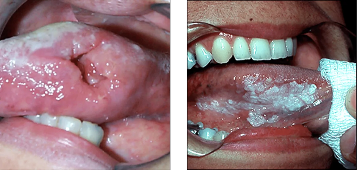
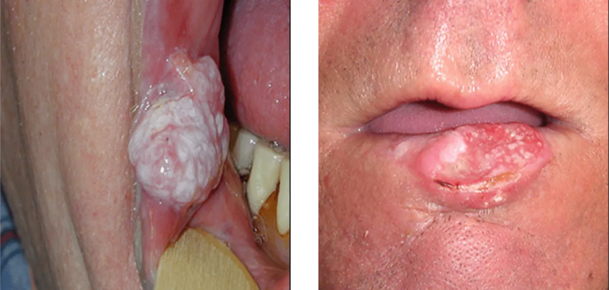
Clinical Signs of Mucosal Pathology and the 14-Day Rule
Oral tissue alterations can occur anywhere within the oral cavity, including the lips, tongue, floor of the mouth, gums, roof of the palate, and buccal cheek mucosa. Common warning signs include non-wipeable white patches (leukoplakia), velvety red patches (erythroplakia), chronic mouth sores that bleed upon contact, painless firm nodules, and localized jawbone expansion. The fundamental clinical benchmark in oral pathology is the 14-day rule: standard aphthous ulcers (canker sores) and traumatic cheek bites heal naturally within ten to fourteen days. Any mucosal sore, ulcer, or lump that persists beyond two weeks warrants immediate professional evaluation by an oral surgeon.
Diagnostic Biopsy Modalities and Histopathologic Analysis
Visual inspection and palpation alone cannot provide a definitive medical diagnosis. An oral biopsy remains the gold standard diagnostic method, involving the gentle removal of a small tissue sample for microscopic histopathologic analysis. Depending on the size, location, and characteristics of the lesion, Dr. Caceres performs either an excisional biopsy or an incisional biopsy. For small, localized nodules such as irritation fibromas or minor salivary gland mucoceles, an excisional biopsy completely removes the lesion along with a tiny rim of healthy tissue, serving as both diagnosis and definitive cure. For larger or diffuse patches, an incisional biopsy samples a representative wedge of tissue.
| Lesion Classification | Clinical Presentation & Causes | Diagnostic Protocol & Action |
|---|---|---|
| Benign Traumatic Lesion | Irritation fibroma, mucocele, or traumatic ulcer from chronic bite irritation | In-office excisional biopsy; curative removal in 15 minutes with dissolvable sutures |
| Autoimmune / Inflammatory | Oral lichen planus with lacy white Wickham striae or burning desquamation | Incisional punch biopsy to confirm histology, followed by topical anti-inflammatory therapy |
| Premalignant Dysplasia | Leukoplakia (white plaque) or erythroplakia (red velvety patch) with cellular changes | Targeted incisional biopsy to grade dysplasia; complete surgical excision if moderate/severe |
| Odontogenic Cysts & Tumors | Dentigerous cyst or ameloblastoma expanding inside the upper or lower jawbone | 3D CBCT imaging, aspiration biopsy, followed by surgical enucleation and bone curettage |
Patient Comfort, In-Office Anesthesia and Chairside Reality
Many patients fear that an oral biopsy is an extensive, painful procedure, but in reality, an in-office mucosal biopsy is brief, gentle, and routinely completed in fifteen to twenty minutes. The target tissue is thoroughly numbed with targeted local anesthetic, ensuring you experience zero pain during tissue sampling. For anxious patients, Dr. Caceres provides nitrous oxide laughing gas or comfortable IV twilight sedation in our surgical suite. Once the sample is retrieved, one or two tiny dissolvable sutures are placed to support rapid closure. You can comfortably drive yourself home and resume normal daily activities the following morning.
Pathology Turnaround, Result Categories and Treatment Protocols
All biopsy specimens are immediately placed in fixative and sent to a board-certified oral and maxillofacial pathology laboratory for microscopic analysis. Histologic results are typically available within five to seven business days. Dr. Caceres personally reviews the pathology report with you, explaining the cell findings in clear, understandable language. If the lesion is benign, no further surgical intervention is necessary. If dysplastic or precancerous changes are detected, Dr. Caceres coordinates appropriate curative surgical excision and regular monitoring protocols, ensuring your oral health is protected at every stage.
Schedule an Oral Pathology Consultation
Do not wait and worry about persistent mouth sores or lumps. Board-certified oral surgeon Dr. Johanny Caceres provides gentle oral pathology evaluations and prompt diagnostic biopsies in Coral Gables.