
The Hidden Problems Wisdom Teeth Can Cause
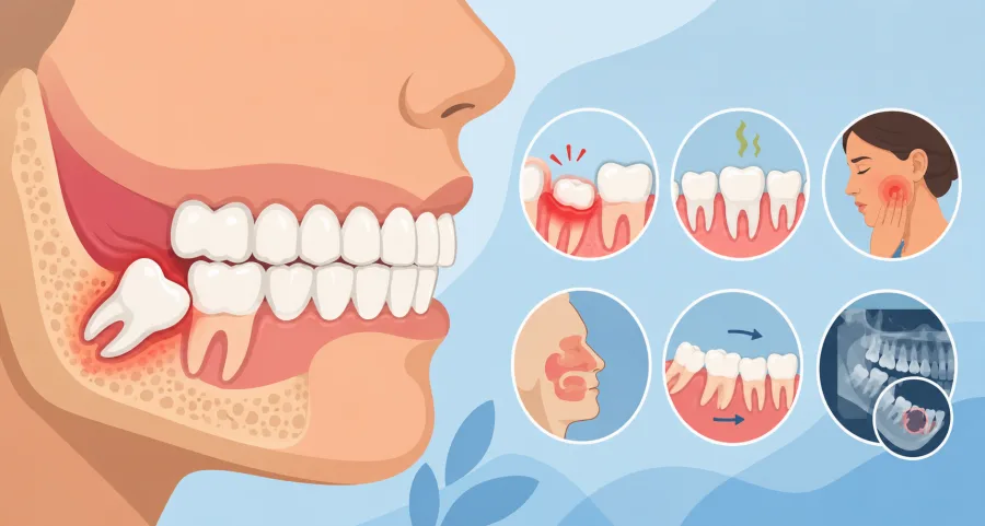
When people consider wisdom teeth, they usually picture visible swelling, sudden throbbing pain, or difficulty opening their mouth. However, some of the most destructive oral surgery conditions develop in complete silence. Impacted and malpositioned third molars can spend years quietly undermining your dental structure, dissolving surrounding jawbone, and damaging healthy adjacent teeth without generating a single ache until the damage is already extensive.
At Oral and Facial Surgery of Miami, board-certified oral and maxillofacial surgeon Dr. Johanny Caceres frequently identifies asymptomatic pathology on routine 3D radiographic scans. Understanding these concealed biological risks illustrates why specialized oral surgeons recommend proactive diagnostic evaluations rather than waiting for an emergency to strike.
Silent Pathology: Odontogenic Cysts and Mandibular Jaw Weakening
Every tooth forms inside a fluid-filled protective envelope of tissue known as the dental follicle. When a wisdom tooth erupts normally, this tissue sac naturally breaks down. However, when a third molar remains completely trapped beneath dense cortical bone, the follicle can continue accumulating fluid and expand into an odontogenic dentigerous cyst.
Dentigerous cysts grow slowly and painlessly over several years. As they expand, they hollow out the internal cancellous framework of the lower jawbone (mandible), eroding the structural integrity of the jaw. In severe cases, an untreated cyst can weaken the jaw so significantly that a minor bump or firm chewing forces can cause a pathological mandibular fracture. In addition, cystic lining cells can occasionally undergo neoplastic transformation into aggressive odontogenic tumors such as ameloblastomas, requiring major reconstructive bone resection.
| Hidden Complication | Pathological Mechanism | Clinical Consequence |
|---|---|---|
| Dentigerous Cyst | Fluid accumulation within un-erupted follicular sac | Bone cavitations, structural jaw thinning, tooth displacement |
| Second Molar Resorption | Chronic physical pressure from mesioangular crown | Dissolution of healthy second molar root cementum and dentin |
| Periodontal Bone Pocket | Anaerobic plaque trapped in interproximal crevice | Permanent bone loss exceeding 7mm behind the terminal molar |
| Maxillary Sinus Infiltration | Upper molar roots penetrating thin sinus floor | Chronic unilateral sinusitis, sinus membrane thickening, congestion |
Structural Destruction to Neighboring Second Molars
The second permanent molar sits directly adjacent to the wisdom tooth and carries immense chewing value. Unfortunately, it frequently becomes the collateral victim of impacted third molars. When a wisdom tooth erupts at a 45-degree or 90-degree horizontal angle, the hard enamel crown pushes directly into the back surface of the second molar root trunk.
This constant biomechanical friction activates osteoclast cells in the periodontal ligament, triggering external root resorption. In essence, the body begins eating away at the root of the healthy tooth. By the time the patient feels a toothache, a massive cavity or crater has hollowed out the second molar below the gumline. In many unfortunate scenarios, oral surgeons must extract both the wisdom tooth and the compromised second molar because the damage cannot be repaired with fillings or crowns.
Upper Wisdom Teeth and Chronic Maxillary Sinusitis
While lower wisdom teeth threaten the mandibular canal and masseter muscles, upper third molars interact closely with your respiratory system. The roots of upper wisdom teeth develop immediately beneath the bony floor of the maxillary sinus, separated by only a paper-thin layer of cortical bone or Schneiderian membrane.
When an upper wisdom tooth becomes impacted or infected, bacteria readily migrate across this delicate sinus floor. Patients often spend years seeing allergists and primary care physicians for recurrent unilateral nasal congestion, sinus pressure beneath the cheekbones, post-nasal drip, and chronic headaches, unaware that an impacted upper molar is driving their respiratory distress. Removing the upper tooth eliminates the chronic bacterial reservoir and allows the sinus cavity to heal permanently.
Diagnostic Detection and Minimally Invasive Removal at OFS Miami
The only reliable way to uncover these hidden conditions before they inflict permanent damage is through high-definition 3D Cone Beam Computed Tomography (CBCT). Unlike flat 2D panoramic X-rays, 3D imaging allows Dr. Johanny Caceres to rotate your anatomy on three distinct spatial planes, inspecting bone density, cyst boundaries, sinus floors, and nerve paths down to fractions of a millimeter.
At our state-of-the-art Coral Gables surgical facility, Dr. Caceres utilizes minimally invasive microsurgical techniques to safely remove impacted third molars and enucleate any cystic tissue. Combined with customized IV sedation, your procedure is comfortable, quick, and stress-free.
Do not wait for quiet symptoms to become loud dental emergencies. Contact Oral and Facial Surgery of Miami today at (305) 552-1193 or schedule your comprehensive surgical consultation online today.