
Wisdom Tooth Nerve Damage: How Rare Is It, Really?
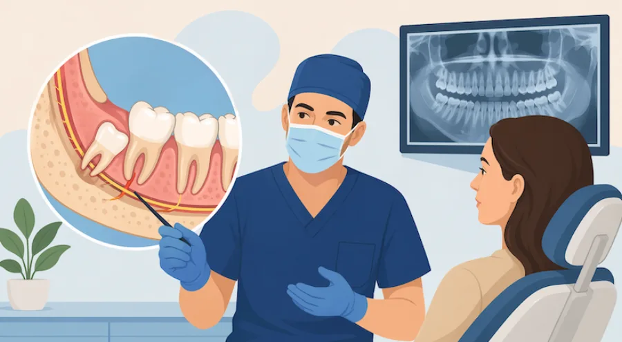
Hearing the words "nerve damage" before a wisdom tooth procedure is enough to give any patient pause. Online discussion forums often amplify rare worst-case complications, leaving many patients feeling anxious about whether extracting their lower third molars could leave their lip or tongue permanently numb. When a dentist or oral surgeon mentions that a wisdom tooth root sits close to a nerve canal, understanding the true clinical risk is vital for peace of mind.
The clinical reality is reassuring: permanent nerve injury from wisdom tooth extraction is extraordinarily rare. By reviewing maxillofacial anatomy, examining the medical classification of nerve responses, and looking at rigorous epidemiological data, patients can replace fear with evidence-based confidence.
Anatomy and Pathways: Mapping the Inferior Alveolar and Lingual Nerves
To understand why nerve proximity is discussed during your consultation, it helps to understand the anatomy of the lower jaw (mandible):
- The Inferior Alveolar Nerve (IAN): This major sensory branch of the mandibular nerve (trigeminal cranial nerve V3) travels through a bony channel called the mandibular canal, situated just beneath or between the roots of the lower molar teeth. The IAN supplies pure sensation (touch, temperature, and pain) to your lower teeth, lower lip, and chin. It controls no facial expression or motor movements, meaning any injury affects feeling, not your smile.
- The Lingual Nerve: This nerve runs along the inner (lingual) soft tissue wall of the lower jaw, adjacent to the tongue-side bone plate near the wisdom tooth. It supplies sensation and taste to the anterior two-thirds of the tongue and the floor of the mouth.
- The Long Buccal Nerve: A minor sensory nerve providing feeling to the small patch of cheek tissue alongside the lower molars. Irritation to this tiny branch rarely causes noticeable symptoms.
Because lower wisdom teeth develop late in adolescence, their roots grow downward toward the mandibular canal. In some individuals, root tips merely touch the outer wall of the canal; in others, the nerve actually passes between divergent roots. Upper wisdom teeth, by contrast, are situated far away from these pathways and carry zero risk of lower lip or tongue numbness.
Degrees of Nerve Response: Medical Classifications Explained
In clinical oral and maxillofacial surgery, nerve disturbances are categorized using the classic Seddon and Sunderland classifications of nerve injury:
- Neuropraxia (Mild Physiological Concussion): By far the most common type of post-operative nerve disturbance. The physical structure of the nerve remains completely intact, but temporary edema or gentle pressure during root elevation causes a temporary conduction block. Patients experience partial numbness, tingling, or an altered pins-and-needles sensation. Sensation typically returns rapidly within days or weeks as localized tissue swelling subsides.
- Axonotmesis (Internal Axonal Disruption): The outer insulating sheath (epineurium) of the nerve remains continuous, but some internal nerve fibers (axons) experience traction damage. The nerve regenerates along its existing anatomical tunnel at a biological rate of roughly one millimeter per day. Full sensory recovery generally takes several months.
- Neurotmesis (Complete Transection): A severe disruption where the nerve trunk is completely severed. This catastrophic event is exceptionally rare in modern oral surgery and typically occurs only during blind surgical instrumentation without 3D imaging.
Real-World Statistics: Temporary Neuropraxia vs. Permanent Injury
When evaluated across large-scale maxillofacial surgical registries, the numbers demonstrate the exceptional safety of modern wisdom tooth extractions:
- Temporary Sensory Disturbance (Neuropraxia): Occurs in approximately 1% to 3% of standard lower third molar extractions, rising to roughly 4% to 8% in deeply impacted cases where roots directly contact the canal.
- Permanent Sensory Alteration: Clinically defined as altered sensation persisting beyond 12 months, permanent nerve damage occurs in less than 0.3% to 0.5% of all cases worldwide.
- Spontaneous Recovery Rate: Over 90% of patients who experience post-operative numbness or tingling experience complete, spontaneous resolution within six to eight weeks without requiring secondary interventions.
It is equally critical to understand the alternative risk: leaving a severely impacted, chronically infected wisdom tooth in place. Chronic pericoronitis, deep periodontal bone loss, and expanding dentigerous cysts erode the mandibular canal from within, exposing the nerve to chronic bacterial toxins and risking permanent pathological nerve compression.
Advanced Prevention and Surgical Precision with Dr. Johanny Caceres in Coral Gables
Minimizing nerve risk begins with specialized diagnostic technology and surgical expertise. Board-certified oral and maxillofacial surgeon Dr. Johanny Caceres utilizes state-of-the-art in-office 3D Cone Beam Computed Tomography (CBCT) at Oral and Facial Surgery of Miami in Coral Gables.
Unlike traditional two-dimensional panoramic X-rays, which flatten 3D structures and make roots appear to overlap the nerve canal, high-resolution CBCT cross-sections reveal the precise spatial distance between the tooth roots and the nerve bundle in sub-millimeter increments.
If a tooth is positioned intimately against the nerve, Dr. Caceres utilizes micro-surgical tooth sectioning, removing the crown in delicate segments to avoid applying hydraulic pressure against the canal floor. In select high-risk anatomical scenarios, a coronectomy (removing the problematic crown while intentionally leaving the healthy root tips buried undisturbed beneath bone) can be performed to reduce nerve risk to virtually zero.
Do not let exaggerated online fears prevent you from protecting your oral health. Contact our Coral Gables surgical team directly at (305) 552-1193 or schedule your consultation online today.