
How Bone Density Affects Wisdom Tooth Removal Surgery
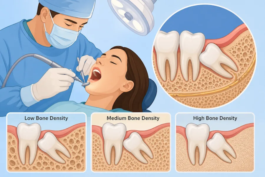
When evaluating wisdom teeth for extraction, most patients focus on whether the teeth are causing pain, crowding their smile, or pushing against neighboring molars. While these symptoms are critical, oral surgeons assess an equally important anatomical factor that dictates the technical difficulty of the procedure: bone density. The structural thickness, mineralization, and elasticity of the jawbone surrounding a third molar directly determine how the tooth is removed and how quickly the socket heals.
At Oral and Facial Surgery of Miami, board-certified oral and maxillofacial surgeon Dr. Johanny Caceres utilizes advanced 3D Cone Beam Computed Tomography (CBCT) to evaluate jawbone architecture before every surgical extraction. Understanding how bone density influences surgery provides valuable clarity on why proactive treatment during youth is so widely recommended.
Biomechanical Foundations: Cortical vs Trabecular Bone Density
Human jawbones are composed of two distinct types of bone tissue: an outer shell of compact, dense cortical bone and an inner core of porous, spongy trabecular (cancellous) bone. The ratio of cortical to trabecular bone varies dramatically between different areas of the mouth. In the upper jaw (maxilla), bone is predominantly cancellous and naturally softer, allowing upper wisdom teeth to be delivered with relatively gentle elevation mechanics.
In contrast, the lower jaw (mandible) features thick, highly calcified cortical plates engineered to withstand powerful chewing forces. Lower wisdom teeth are frequently encased in dense cortical bone known as the external oblique ridge. When bone density is exceptionally high, the bone has zero flexibility, meaning the tooth cannot be nudged or luxated without removing restrictive bone or dividing the tooth into smaller segments.
| Bone Density Type | Anatomical Location | Surgical Approach Required |
|---|---|---|
| Type I (Dense Cortical) | Anterior and posterior adult mandible | Copious saline irrigation, surgical troughing, tooth sectioning |
| Type II (Porous Cortical / Dense Trabecular) | Young adult mandible and posterior maxilla | Targeted osteotomy, moderate luxation, rapid vascular healing |
| Type III (Porous Trabecular) | Standard maxillary tuberosity | Conservative elevation protecting sinus floor and tuberosity bone |
| Type IV (Fine Trabecular) | Elderly or osteopenic posterior maxilla | Delicate instrument handling to prevent sinus membrane perforation |
Why Age Dramatically Changes Bone Density and Surgical Mechanics
The relationship between age and bone density is the primary biological reason why oral surgeons advocate for wisdom tooth evaluation during late adolescence. In teenagers and individuals in their early twenties, jawbone has not yet completed full calcification. The bone is more vascular, contains larger marrow spaces, and possesses natural elasticity that gives slightly under gentle surgical pressure.
As individuals progress into their late twenties, thirties, and forties, bone mineral density peaks. The alveolar bone becomes rigid, brittle, and tightly fused to the cementum of tooth roots. If an adult requires wisdom tooth extraction, the surgeon cannot rely on bone flexibility. Attempting to force a tooth from dense bone without sectioning risks fracturing root tips or placing undue stress on the mandibular jaw. Sectioning the tooth into independent segments allows each piece to be removed cleanly along its individual path of exit.
Surgical Techniques for High-Density Impactions
Managing high-density bone requires meticulous surgical precision and specialized instruments to safeguard adjacent tissues:
- Copious Saline Irrigation: When bone drills or piezosurgery instruments trim dense bone, friction generates heat. Continuous chilled saline irrigation prevents thermal bone necrosis, ensuring the surrounding bone cells remain healthy and capable of rapid healing.
- Multi-Planar Tooth Sectioning: Rather than expanding dense bone, Dr. Caceres divides the wisdom tooth into precise halves or quadrants, lifting each root independently to preserve the surrounding bone socket.
- Coronectomy Protocols: In adult patients where hyperdense bone anchors curved roots in direct contact with the inferior alveolar nerve, Dr. Caceres may recommend a coronectomy. This specialized technique removes the crown while leaving undisturbed root tips safely embedded, entirely avoiding nerve trauma.
- Site Preservation Grafting: In cases where substantial bone removal was required to deliver a deeply impacted tooth, bone graft biomaterials can be placed into the socket to rebuild alveolar volume and support the neighboring second molar.
Precision Diagnostics and Recovery Management at OFS Miami
At our practice, we eliminate procedural uncertainty through high-resolution 3D CBCT imaging. This advanced technology maps cortical bone thickness, marrow spacing, root divergence, and nerve canals in sub-millimeter detail before surgery begins.
To ensure absolute comfort during surgery regardless of bone density, Dr. Caceres provides customized outpatient IV sedation in our accredited Coral Gables surgical suite. You sleep comfortably while your surgical team completes the procedure safely and efficiently.
Discover how personalized surgical planning ensures a smooth extraction and gentle recovery. Contact Oral and Facial Surgery of Miami today at (305) 552-1193 or reserve your consultation online today.